Knee Joint Anatomy: A Comprehensive Guide
Introduction
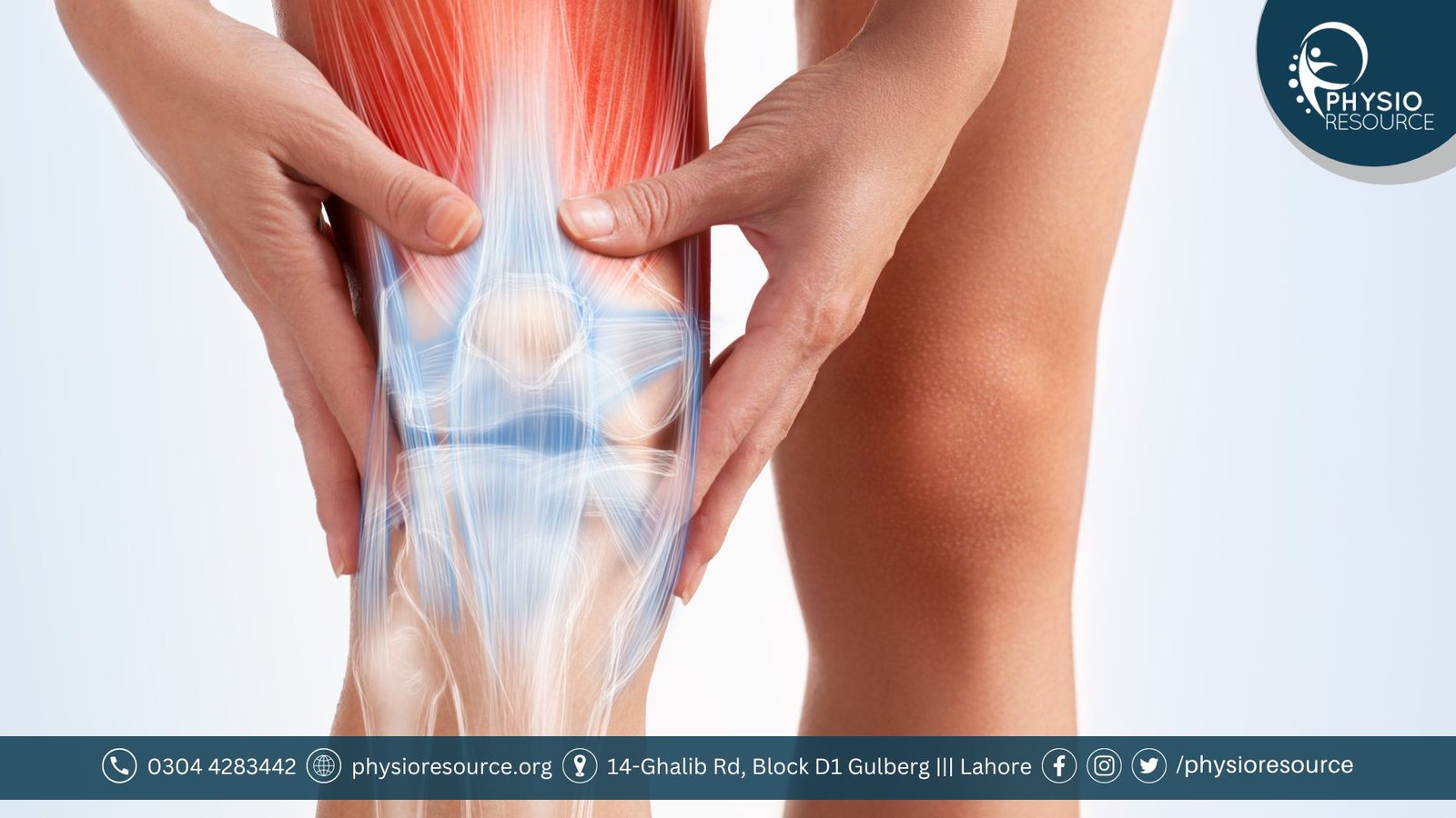
In human body the most complex and the largest joint is the knee joint. This complex knee joint structure is made up of muscles, four bones and ligaments complex web. Knee joint is a bi-condylar (hinge) form of synovial joint, due to this it can perform flexion and extension and little degree of medial and lateral rotation. Knee joint is the most stressed joint of the body.
Anatomy
Articulating surfaces
In knee joint the femur, tibia and patella articulates through 2 joints which are patellofemoral and tibiofemoral joints. Articular cartilage helps to reduce friction forces between these three bones because they are very frim and smooth.
The articulation between the patella and the femur’s anterior and posterior portion form the
patellofemoral joint. The patella is located in the intercondylar groove of femur.
Similar to this, the articulation between medial and lateral condyles of femur and tibia’s condyle form a tibiofemoral joint. Which is the weight bearing joint of the knee.
The tibiofibular joint is also present there, which is formed between tibia and fibula. Although this joint is not involved directly in knee joint, but it is important for the attachment of muscles and ligaments.
Menisci
In knee joint medial and lateral menisci are present which crescent shaped fibrocartilage rings are. The surface of each menisci is concave superiorly and flat inferiorly providing the surfaces to femoral condyles and tibial plateau respectively. The both menisci are connected at both ends of tibia’s intercondylar region. The menisci has the 2 main functions.
- The tibiofemoral joint stability is increased because menisci deepens the surface of tibia.
- Menisci serve as shock absorbers due to increase in area of contact and improve the weight distribution across the joint.
The meniscal fibers arrangement distributes axial loads radially, which decreases the hyaline cartilage’s wear. Which is important during walking or running, because during walk knee stresses can increase to 1-2 times and 3–4 times the body weight during running.
The medial meniscus are less mobile because they are fixed with the joint capsule and medical collateral ligament. Therefore due to damage of medial collateral ligament, the medial meniscus tears occurs frequently.
The lateral meniscus is more mobile and smaller than the medial meniscus because it does not have any additional attachments.
The both menisci’s are fully vascularized during 1st year of life. But after human starts weight bearing the vascularity in meniscus decreases in the red zone (outer 3rd part). While the synovial fluid provides nutrition to the white zone (non-vascularized part) through diffusion.
Also Read: Best Workout Split for Muscle Gain
Bursa
There are 4 synovial fluid filled sacs are present between the moving parts of knee joint known as bursa. Which are semimembranosus, suprapatellar, infrapatellar and prepatellar bursa.
The function of bursas to decrease wear and tear on those knee joint structures.
Joint Capsule
Joint capsule enhance the joint stability. It limits the joint movements and provide the passive joint stability and it has proprioceptive nerve endings due to which active joint stability increases.
Joint capsule has a membrane called synovial membrane and this membrane produces synovial fluid which provide nourishment to the surrounding structures of knee joint.
Ligaments
Each ligament of knee joint provide the optimal stability to knee joint and each has particular function. Knee has two main types of ligaments.
Collateral Ligaments
They are present on the both opposite (medial and lateral) sides of knee in strap-like form.
- Medial collateral ligament (MCL): This ligament attached proximally to the medial epicondyle of the femur and distally with the medial condyle of tibia. It helps to prevent excessive medial movement of knee joint. It resists the valgus force (force from the outside of knee) more effectively during full knee extension (closed packed position) because there is more laxity in ligament during knee flexion (open packed position).
- Lateral collateral ligament (LCL): It is an extracapsular ligament. This ligament is attached with the lateral epicondyle of femur proximally and with lateral side of fibula head distally. It helps to prevent excessive lateral movement of knee joint. It resists the varus force more effectively.
Cruciate Ligaments
Tibia and fibula are connected by the 2 cruciate ligaments and they create an X when they cross each other in knee joint.
- Anterior cruciate ligament (ACL): Anterior dislocation of tibia on femur is prevented through this ligament. It is attached tibia’s anterior intercondylar region with femur’s intercondylar fossa.
- Posterior Cruciate Ligament (PCL): This ligament attach the tibia’s posterior intercondylar region with femur’s anteromedial condyle. PCL helps to stops tibia from dislocating on femur posteriorly.
Blood and Nerve Supply
The main Arteries are involved in the knee joint are:
The Nerves in the knee joint are:
Muscles and Functions
These 4 movements of knee joint performed by the following muscles.
FLEXION:
- Hamstrings (Biceps femoris, Semitendinosus, Semimembranosus)
EXTENSION:
- Quadriceps femoris (vastus lateralis, rectus femoris, vastus intermedius and vastus medialis)
- Tensor fasciae latae ( weak Extensor)
Medial Rotation:
- Sartorius
- popliteus
- gracilis
- semitendinosus
- Semimembranosus
Lateral Rotation:
Common Injuries and Conditions of Knee Joint
- Knee osteoarthritis
- Patellofemoral pain syndrome (PAS)
- Medial collateral ligament (MCL) injury
- Lateral collateral ligament (LCL) injury
- Posterior cruciate ligament (PCL) injury
- Anterior cruciate ligament (ACL) injury
- Quadriceps muscle contusion
- Sinding Larsen Johansson syndrome
- Osgood-Schlitter’s disease
- Articular cartilage lesions
- Osteochondritis dissecans of the knee
- Quadriceps muscle contusion
- Semimembranosus tendinopathy
Common Symptoms of Knee Joint Disorder
Following are the symptoms of knee joint disorder that you need to contact medical professionals or a physiotherapist:
- Severe pain around knee joint.
- Popping sound from knee joint during walking, stair climbing and moving the leg.
- Unable to bear weight on knee
- Stiffness around knee joint
- Swelling
- Warmth in contact
- Redness
- Weakness
- Unable to straight and bend knee joint
- Difficulty in climbing up the stairs
- Knee joint locking and buckling
- Even after three days of at-home therapy, your pain persists.
- Below the aching knee, you get calf discomfort, numbness, swelling, tingling, or bluish discoloration.
- Knee has a malformation or deformity, for example Varus or Valgus deformity
Knee Pain Treatment in Lahore
Visit us at Physio Resource if you are unable to enjoy life fully due to knee pain. Knee is the most important joint of human body which help us move and walk. Our physiotherapist will find therapies that maintain your knees strong, healthy, and functioning as they should as well as assist you in understanding what’s happening inside your knee joint. They are dedicated to provide everyone with high-quality physiotherapy care through state-of-the-art facilities and highly qualified professionals. So scheduled your appointment now with one of our best physiotherapist in Lahore.
Contact us
Phone No: 0304-4283442
Address: 14-Ghalib Rd, Block D1, Gullberg III, Lahore


Najma Batool
July 27, 2024This is an excellent, thorough guide about the knee joint! Finding out how everything functions in harmony to keep us going is incredible.